Forschung
MS Imaging of food - molecular specificity and spatial information
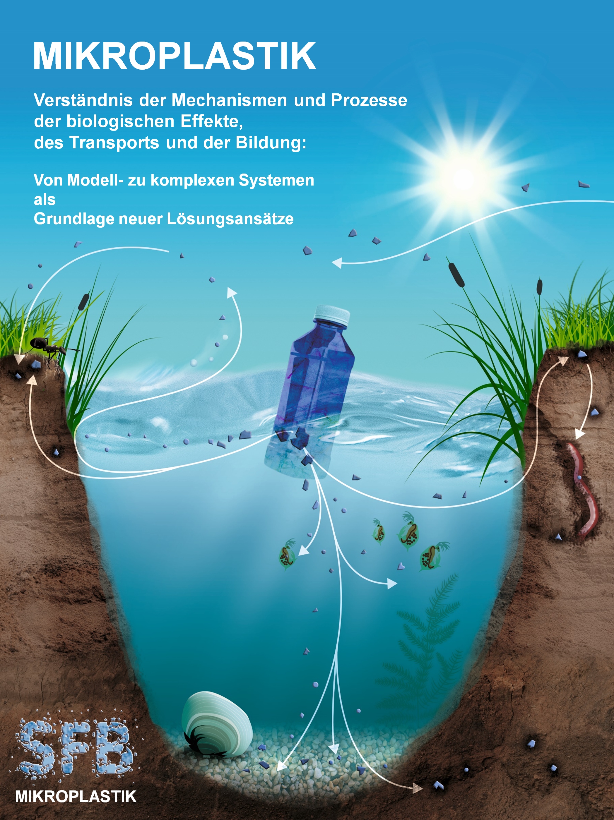
The ubiquitous presence of microplastic (MP) particles has been documented in oceans, land, freshwater systems, air and drinking water. MP particles are commonly referred to as particles with a diameter < 5 mm and are thought to be readily taken up by organisms through inhalation or ingestion. This has raised concerns about possible health impacts on living organisms including humans. The question if MP particles cause effects by purely passing through the intestinal tract or only after translocation into the tissue is not fully understood yet. It is also not clear which properties of MP particles are responsible for these effects. Therefore, this project aims at directly correlating local molecular and histological effects in tissue to individual particles. This will be achieved by a combination of spatially resolved analysis methods and classical histology. The experiments will be based on MP particles of different composition and morphology (spherical particles, fragments, fibers) and aquatic and terrestrial model organisms. The analysis of the same tissue section by highly complementary methods will enable us to relate molecular and histological effects in tissue directly to a single (identified) MP particle and will thus provide new insight into processes causing MP-related effects. This work is part of the Collaborative research Center ‘Microplastics’ (SFB 1357) at the University of Bayreuth.
Latest publications:
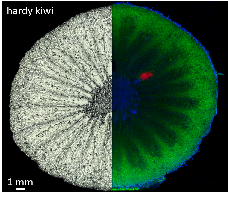
MS Imaging allows visualising the distribution of chemical components throughout the food matrix. This can be of interest when analytes are expected to be located only in particular tissue parts, e.g. only in the mesocarp or in the peel of a fruit. This project focuses on MS Imaging of processed food. Especially in processed food consisting of several ingredients, the distribution not only of endogenous compounds, such as nutrients, but also of food additives, nutrients for food fortification, contaminants or other minor components are of interest and more challenging to predict. Current work deals with the penetration of preservatives into cheese in the context of official penetration limits defined by European legislation and the investigation of nutrients’ and particularly contaminants’ distributions in processed food of plant origin in the light of mitigation methods for the reduction of contaminants in food.
Latest Publications:
Microplastics - Effect on Organisms - SFB 1357
Evaluation of new antibiotics by MS imaging - DZIF
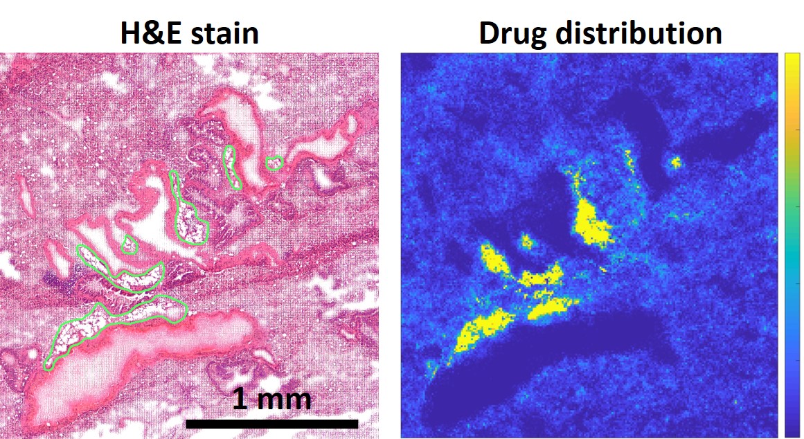
Tuberculosis (TB) is an infectious disease of the lungs caused by Mycobacterium tuberculosis. TB, which is generally considered to be eradicated in developed countries, is still very common in developing countries in Africa and Asia and according to the WHO remains the leading cause of death from an infectious disease before HIV/AIDS, Malaria and Lepra. Recent years have shown an increase in the prevalence of drug resistant TB strains creating an increasing demand for novel anti-TB drugs. As part of the German Center for Infection Research (DZIF), we work on developing a universal platform to test and evaluate the in vivo efficacy of novel anti-TB drugs. To this end, we employ a specialised technique called MALDI mass spectrometry imaging (MALDI-MSI), which is able to visualise the distribution of drug compounds, drug metabolites and other biomolecules in tissue samples. In our studies, we apply MALDI-MSI to investigate the distribution of TB antibiotics in tissue samples of a novel mouse model, which closely resembles human TB pathology. Through this, we aim to reach credible predictions about a drug's in vivo efficacy, before it enters clinical trials. In close cooperation with the Institute of Tropical Medicine of the LMU Munich as well as the Leibniz Lung Center, we aim to accelerate the development of novel TB antibiotics to meet this global health issue.
Latest publications:
Origin and authenticity of cereals by Raman spectroscopy - AgrOr
Grain is an important staple food and is subject to substitution and adulteration. Currently, grain is the 5th most commonly counterfeited food in the world. Therefore, origin and authenticity are important. In this cooperative project a combination of spectroscopic methods (NMR, Raman, NIR and MIR) and isotope ratio mass spectrometry is applied to verify origin and authenticity of cereals. To this end, spectral patterns and compounds are sought which can be used as fingerprint and markers for origin, species, purity of variety and cultivation method.
Raman spectroscopy is used as a non-invasive method which requires minimal sample preparation with a view to an application for inspection of commodities. Raman spectra are providing - as a fingerprint - information about molecular vibrations and thus the composition of the samples. The Raman subproject specifically aims at identifying species, variety, adulteration and origin using confocal Raman mapping with multivariate data analysis. Thus, the fingerprint spectra are used for discrimination and also for identification of components (for example starch, proteins, ferulic acid…).
imzML - open data format for mass spectrometry imaging - www.imzml.org
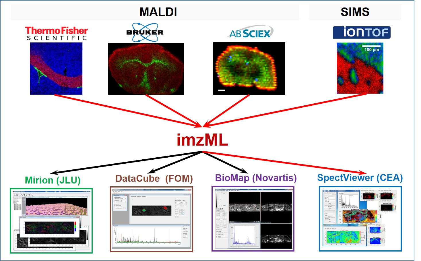
Open data formats are key to facilitating data sharing, integration and interoperability. imzML is an open, vendor-neutral format for mass spectrometry imaging data [1]. It provides more flexibility in data processing as the user is no longer limited to proprietary software but can choose the processing option that best fits the purpose. Measurement results (full data sets) can be shared between collaborators without loss of information (which is the case when sharing only the images). imzML is thus an important step towards more efficient collaboration as well as towards more transparent reporting of results in mass spectrometry imaging. All relevant information on imzML including technical specifications, available software tools and implementation examples are provided on the website www.imzml.org. The range of MS imaging software that supports imzML is constantly growing (see website for a complete list). We have recently developed a new imzML validation and editor tool to enable users to ensure that any given imzML file is valid according to the specification and will therefore function correctly in the wide variety of available tools supporting imzML.
Validator for imzML (and mzML)
Error-Free Data Visualization and Processing through imzML and mzML Validation.
Multicenter study (data integration based on imzML)
Mass spectrometry imaging of biological tissue : an approach for multicenter studies.
'Open data' for imzML
A public repository for mass spectrometry imaging data.
imzML specifications
imzML: a common data format for the flexible exchange and processing of mass spectrometry imaging data.
Mobile Raman Analysis
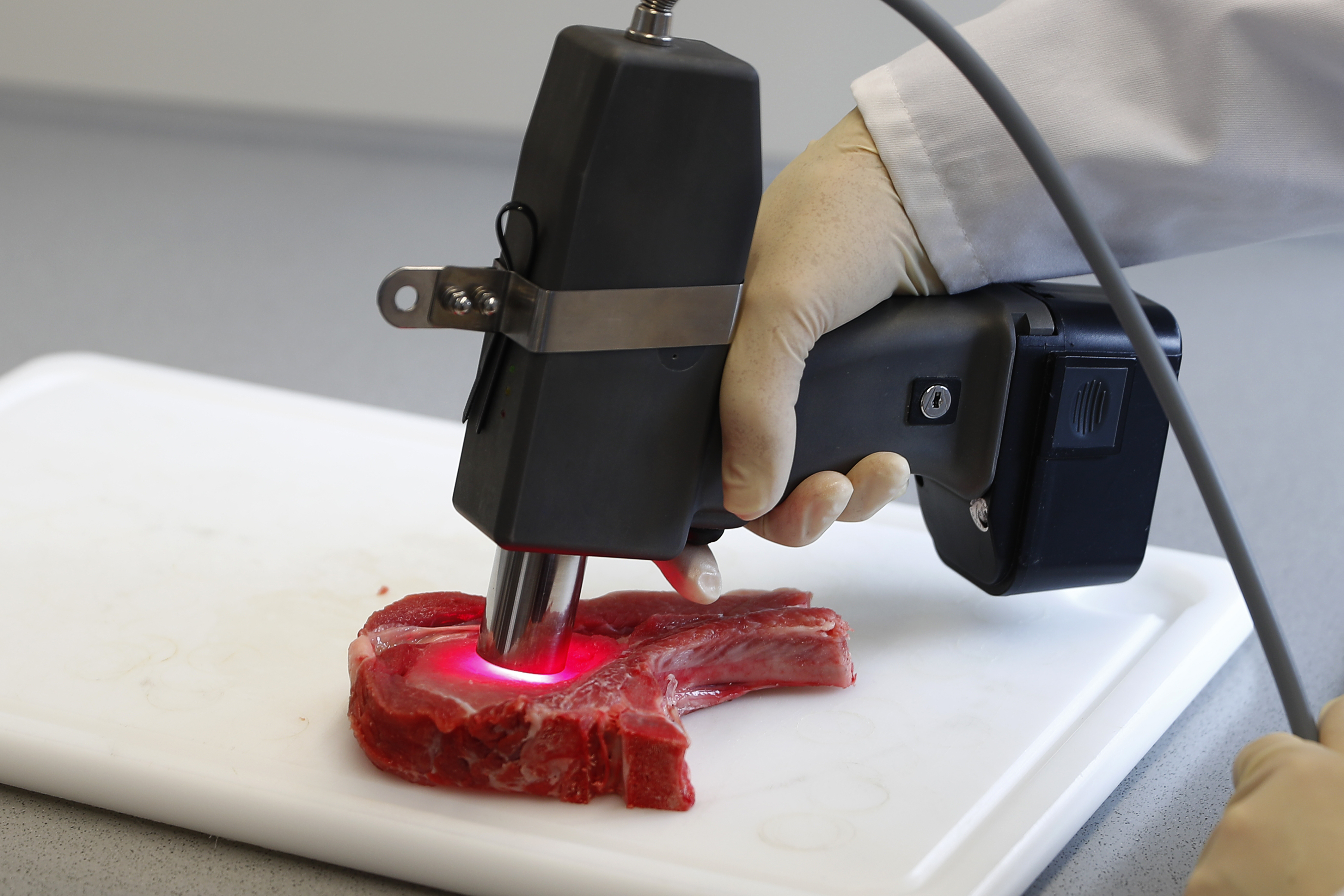
Raman spectroscopy is used as a non-invasive and non-destructive measuring method to investigate the quality of raw materials and food on site. This is of interest for the inspection of incoming goods, for sorting and quality management. To this end, two mobile Raman scanner have been developed which allow for the objective detection or a prediction of quality traits such as pH, drip loss or tenderness of meat in an abattoir or during processing. The scattered light provides information about the molecular composition. Thus, biochemical, structural and microbiological changes can be detected.